Hip Arthroscopy & Preservation
Hip Arthroscopy
What is Hip Arthroscopy?
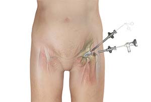
Arthroscopy, also referred to as keyhole or minimally invasive surgery, is a procedure in which an arthroscope is inserted into a joint to check for any damage and repair it simultaneously.
An arthroscope is a small, fiber-optic instrument consisting of a lens, light source, and video camera. The camera projects an image of the inside of the joint onto a large screen monitor allowing the surgeon to look for any damage, assess the type of injury, and repair the problem.
Indications for Hip Arthroscopy
Hip arthroscopy is a surgical procedure performed through very small incisions to diagnose and treat various hip conditions including:
- Removal of torn cartilage or bone chips that cause hip pain and immobility.
- Repair a torn labrum: The labrum is a fibrous cartilage ring which lines the acetabular socket.
- Removal of bone spurs or extra bone growths caused by arthritis or an injury.
- Removal of part of the inflamed synovium (lining of the joint) in patients with inflammatory arthritis. This procedure is called a partial synovectomy.
- Repair of fractures or torn ligaments caused by trauma.
- Evaluation and diagnosis of conditions with unexplained pain, swelling, or stiffness in the hip that does not respond to conservative treatment.
Procedure of Hip Arthroscopy
Hip arthroscopy is performed under regional or general anesthesia depending on you and your surgeon's preference.
Your surgeon will make 2 or 3 small incisions about 1/4 inches in length around the hip joint. Through one of the incisions an arthroscope is inserted. Along with it, a sterile solution is pumped into the joint to expand the joint area and create room for the surgeon to work.
The larger image on the television monitor allows the surgeon to visualize the joint directly to determine the extent of damage so that it can be surgically treated.
Surgical instruments will be inserted through other tiny incisions to treat the problem.
After the surgery, the incisions are closed and covered with a bandage.
Advantages of Hip Arthroscopy
The advantages of hip arthroscopy over the traditional open hip surgery include:
- Smaller incisions
- Minimal trauma to surrounding ligaments, muscles, and tissues
- Less pain
- Faster recovery
- Lower infection rate
- Less scarring
- Earlier mobilization
- Shorter hospital stay
Risks and Complications of Hip Arthroscopy
As with any surgery, there are potential risks and complications involved. It is very important that you are informed of these risks before you decide to proceed with hip arthroscopy surgery. Possible risks and complications include:
- Infection at the surgical incision site or in the joint space
- Nerve damage which may cause numbness, tingling, pain, and weakness
- Excess bleeding into the joint, a condition called hemarthrosis
- Blood clots may form inside the deep veins of the legs which can travel to the lungs (pulmonary embolism).
Postoperative Care following Hip Arthroscopy
Your doctor may advise you to take certain precautions to promote faster recovery and prevent further complications. These include:
- Taking pain medications as prescribed.
- Use of crutches to prevent or limit bearing weight on the operated hip
- Physical therapy exercises should be performed to restore normal hip function and improve flexibility and strength
- Eating a healthy diet and avoiding smoking will help in faster healing and recovery
- Avoid activity which involves lifting heavy things or strenuous exercises for first few weeks after surgery
With advances in surgical techniques, arthroscopy plays an important role in diagnosis and Treatments for hip diseases. Also, patients can anticipate a quicker recovery with less postoperative complications following hip arthroscopy surgery.
Hip Preservation
Hip Anatomy
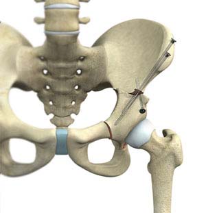
The hip is a ball and socket joint comprising of the femur (thighbone) and the pelvic bone. The head of the femur (ball) articulates with a cavity (socket) called the acetabulum in the pelvic bone. To facilitate smooth and frictionless movement of the hip joint, the articulating surfaces of the femur head and acetabulum are covered by spongy articular cartilage. Injury, wear-and-tear and certain diseases can result in the wearing away of the cartilage tissue, causing painful rubbing of bones.
Hip replacement surgeries have long been the choice of treatment, where the damaged parts of the joint are removed and replaced with prosthesis. However, in young active patients, the prostheses are highly prone to wear-and-tear, and the need for repeat surgery. Hip preservation is a surgery that overcomes the limitations of joint replacement.
Indications of Hip Preservation Surgery
Some of the conditions indicated for hip preservation surgery include:
- Femoroacetabular impingement (FAI): friction in the hip joint from abnormal bony irregularities
- Hip dislocation: head of the femur moves out of the socket
- Hip dysplasia: congenital hip condition characterized by a shallow acetabulum
- Labral tear: tear or separation of the labrum, a cartilaginous ring that surrounds the socket and seals the hip joint
- Avascular necrosis: disrupted blood flow to the hip joint, causing death of bone tissue
Hip Preservation Surgery Procedure
Hip preservation surgery includes various techniques:
- Periacetabular osteotomy: Periacetabular osteotomy is a surgical procedure to treat hip dysplasia. This involves cutting the acetabulum from the pelvic bone and repositioning it with screws to allow for a better fit of the femoral head. The procedure reduces pain, restores function and prevents further deterioration of the hip joint, thereby increasing the life of the hip joint and postponing total hip replacement.
- Surgical hip dislocation: Surgical hip dislocation is a surgical technique that involves the dislocation of the hip joint during surgery to facilitate easy access to the inside tissues of the hip joint. It helps your surgeon to clearly view and treat abnormalities present deep into the hip joint.
- Femoral osteotomy: An osteotomy is a surgical procedure that involves cutting and reshaping of a bone. The femur is cut at the end close to the hip joint and realigned so that it forms a normal angle. This improves the distribution of force placed on the joint and prevents wear-and-tear of the cartilage.
- Hip arthroscopy: Arthroscopy, also referred to as keyhole or minimally invasive surgery, is a procedure in which an arthroscope is inserted into a joint to check for any damage and repair it simultaneously. Hip arthroscopy is a surgical procedure performed through very small incisions to diagnose and treat various hip conditions.
The various hip preservation surgeries for severe hip pain and dysfunction in young and active patients have been found to be beneficial and avoid or delay the need for hip replacement surgery.