
Kinomatic Surgical Approach
Personalized Surgical Planning Before You Enter the Operating Room
Every hip and knee is different. Kinomatic uses advanced CT imaging, 3D modeling and virtual reality to help create a surgical plan based on the individual anatomy of each patient.
Before surgery, detailed imaging is transformed into a three-dimensional model of the hip, knee and lower extremity. This allows the surgical team to evaluate alignment, anatomy, implant size and positioning before the procedure begins.
How Kinomatic Surgical Planning Works
- CT-Based Imaging
The process begins with a high-resolution CT scan of the lower extremities. The scan captures detailed information about the shape and alignment of the bones from the hip through the knee and ankle.
Purpose:
To create an accurate digital representation of your individual anatomy.
- Your Anatomy Recreated in 3D
Advanced software converts the CT images into a three-dimensional model of your bones. Rather than planning from a standard anatomical model, the surgical plan is developed using the patient’s own anatomy.
Purpose:
To allow the joint and surrounding alignment to be evaluated from multiple perspectives.
- Detailed Anatomical Analysis
Biomedical engineers analyze the 3D model and identify important anatomical landmarks and measurements.
This includes evaluating the relationship between the hip, knee and ankle and understanding the patient’s individual lower-limb alignment.
Purpose:
To provide detailed anatomical information that supports surgical planning.
- Patient-Specific Implant Planning
Using the 3D model, different implant sizes, positions and orientations can be evaluated before surgery.
The goal is to develop a plan that takes into account the patient’s anatomy as well as the implant system selected by the surgeon.
Purpose:
To plan implant size and positioning before entering the operating room.
- Virtual Reality Surgical Review
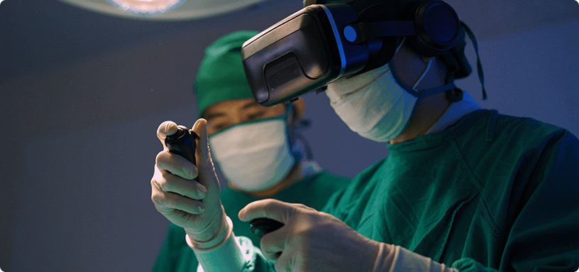
Once the preliminary plan is complete, the surgeon can review the patient’s anatomy and proposed surgical plan in an immersive virtual reality environment.
The 3D model can be rotated and examined from different angles, helping the surgeon review anatomical relationships and make adjustments to the plan when appropriate.
Purpose:
To provide a detailed 360° view of the planned procedure before surgery.
- Surgeon Review and Surgery
The final plan acts as a detailed surgical guide.
Kinomatic technology supports planning, but it does not replace the surgeon’s judgment. Your surgeon remains responsible for the procedure and can make adjustments based on the anatomy and findings encountered during surgery.
Purpose:
From CT Scan to Surgical Plan
Your lower extremity is digitally captured.
Your anatomy is recreated as an interactive three-dimensional model.
Alignment & anatomical measurements are evaluated.
Implant size, position & orientation are considered.
Your surgeon reviews the procedure in an immersive 3D environment.
The patient-specific plan helps guide the procedure.
Why Use 3D & Virtual Reality Surgical Planning?
- Planning Based on Your Anatomy
The surgical plan begins with a model created from your own CT scan rather than relying solely on generalized anatomical measurements.
- A 360° View of the Joint
Three-dimensional visualization allows the surgeon to inspect the hip or knee from multiple angles before surgery.
- Implant Planning Before Surgery
Implant sizing, positioning and orientation can be evaluated as part of the preoperative planning process.
- Detailed Alignment Analysis
The relationship between the hip, knee and ankle can be assessed to better understand the patient’s individual lower-limb alignment.
- Surgeon-Controlled Technology
Virtual reality and 3D planning provide additional information to the surgeon. They do not perform the surgery or replace surgical decision-making.
The Role of Virtual Reality
Seeing More Before Surgery Begins
Traditional X-rays provide valuable two-dimensional information about a joint. CT-based 3D planning adds another perspective by recreating the patient’s anatomy in three dimensions.
Using a virtual reality headset, the surgeon can interact with this model, rotate the joint, examine it from different viewpoints and review the proposed implant position before surgery.
This additional visualization can help the surgeon enter the operating room with a detailed understanding of the planned procedure.
Personalized Planning for Hip & Knee Replacement
Kinomatic surgical planning may be incorporated into:
Hip Replacement
3D planning can help evaluate hip anatomy, implant sizing, component orientation and the relationship between both lower extremities.
Knee Replacement
The surgical plan can incorporate individual knee anatomy, hip-knee-ankle alignment, implant sizing and proposed component positioning.
Technology That Supports the Surgeon
Advanced Planning. Individual Anatomy. Surgeon-Led Care.
Kinomatic combines advanced imaging, biomedical engineering, 3D modeling and virtual reality to provide surgeons with additional information before joint replacement surgery.
The technology helps support preparation and planning, while your surgeon remains responsible for determining the appropriate treatment and performing the procedure.